What Are Surgical Plates and How Do They Work?
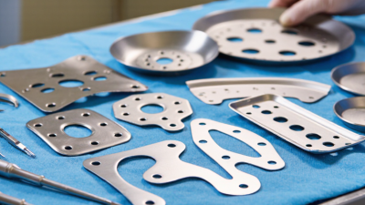
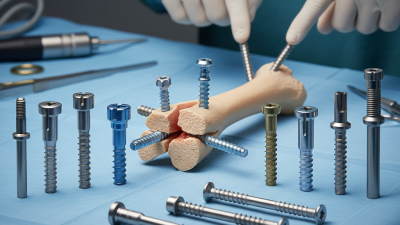
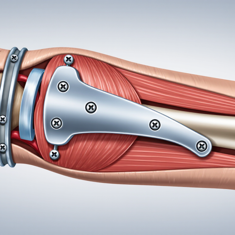
Surgical plates are engineered supports used to stabilize broken bones while natural healing takes place. They sit against the bone and connect across a fracture with carefully selected screws. In practical terms, the plate acts like an internal splint. It helps maintain alignment when a patient walks, lifts an arm, or changes position.
Orthopaedic trauma surgeon Dr. Jesse B. Jupiter has said, “The plate is only one part of the fracture treatment; the biology and the patient matter just as much.” This principle explains why surgical plates are not simple metal patches. Their shape, thickness, screw pattern, and placement must match the fracture and the surrounding soft tissue. Some plates provide compression between bone fragments. Others bridge unstable areas and preserve the fracture’s blood supply. Locking plates can also create a fixed-angle connection, which may help in weaker bone.
The material matters too. Stainless steel and titanium are common choices, but each has different strength, flexibility, and imaging characteristics. A surgeon may select a contoured plate for the wrist, a long bridge plate for the thigh, or a smaller plate for the face. The decision is never automatic. Patient age, bone quality, fracture location, skin condition, and daily activities all influence the plan.
A simple explanation can mislead.
Surgical plates do not repair bone alone. They create conditions for repair. Healing still depends on blood flow, stability, nutrition, rehabilitation, and time. Even experienced surgeons must reassess their assumptions, because a technically secure plate can still fail when biology or patient needs are overlooked.
What Surgical Plates Are: Materials, Types, and ISO 5832 Standards
What Are Surgical Plates and How Do They Work?
What Surgical Plates Are: Materials, Types, and ISO 5832 Standards
Surgical plates are internal fixation devices that support broken or weakened bones. Surgeons place them along the bone and secure them with screws. The plate helps maintain alignment while healing tissue gradually carries more load. It is not a replacement for bone.
Common materials include implant-grade stainless steel, titanium, and cobalt-based alloys. Each offers different strength, flexibility, corrosion resistance, and imaging behavior. Titanium is relatively light and often produces fewer imaging artifacts. Stainless steel can provide high strength and predictable handling. No material is perfect.
Plate design also varies with the injury. Compression plates press fracture surfaces together. Locking plates connect screws to threaded holes, creating a more stable angular construct. Reconstruction plates can be contoured to fit irregular anatomy. A surgeon may choose a narrow, broad, straight, or pre-shaped plate based on bone size and fracture pattern.
ISO 5832 standards define requirements for several metallic implant materials, including chemical composition, mechanical properties, and testing criteria. These standards help confirm that a material meets controlled specifications. They do not guarantee successful healing or eliminate surgical risk. Manufacturing quality, sterilization, plate placement, screw selection, and patient activity all matter.
A plate may appear secure during surgery yet experience changing forces afterward. That detail is easy to underestimate. Bone quality, smoking, infection, and excessive loading can affect outcomes. Material selection therefore requires clinical judgment, imaging review, and careful follow-up rather than reliance on a standard alone.
How Plates Stabilize Fractures Under AO’s Four Fixation Principles
Surgical plates are contoured metal supports placed beside a broken bone. They connect bone fragments with screws and help control movement during healing. A plate does not replace the bone’s natural strength. It shares loads while new bone gradually forms.
The fixation plan follows four connected principles. The first is reduction, meaning the surgeon restores alignment, length, and rotation as closely as possible. Stable fixation then limits harmful movement at the fracture site. Stability may come from compression, a buttress effect, or a bridge construct that spans shattered bone. The correct choice depends on the fracture pattern, bone quality, and soft-tissue condition. No single plate method fits every injury.
Blood supply must also be protected. Excessive stripping, heat, or pressure can damage fragile tissues around the fracture. Surgeons often preserve the surrounding clot and handle bone fragments carefully. Biology matters. Early mobilization is the fourth principle. Once fixation is sufficiently stable, guided movement can reduce stiffness and muscle loss. A patient may begin gentle exercises before full weight-bearing is safe.
A neat X-ray does not guarantee complete healing. Pain, swelling, smoking, poor nutrition, and infection can alter recovery. Follow-up imaging and clinical examinations remain essential. Plate fixation is a balance, not a shortcut. Even experienced teams must reassess the plan as healing develops.
Surgical Placement: Reduction, Drilling, Torque, and Imaging Steps
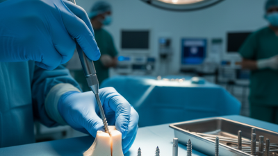
Surgical plates stabilize fractured bone while healing progresses. Placement begins with reduction, restoring length, alignment, and rotation. I look for the joint surface, not only the X-ray. A small rotational error can change gait or hand function.
Once reduction is held, the surgeon marks safe drilling corridors and protects nearby nerves, vessels, and tendons. Drill depth matters. A screw that barely crosses the far cortex may lose purchase; one that projects too far can irritate soft tissue.
Torque is equally practical. Controlled tightening seats the screw without crushing weak bone or stripping its threads. The correct feel is learned through training, tactile feedback, and the implant’s instructions for use. It is not a number to guess.
Imaging checks the work at several points. Fluoroscopy can confirm alignment, screw length, plate position, and joint clearance before closure. The WHO has reported about 234 million major operations worldwide each year, showing why repeatable safety steps matter (Weiser et al., The Lancet, 2008).
In the WHO checklist study, major complications fell from 11.0% to 7.0%, while inpatient deaths declined from 1.5% to 0.8% (Haynes et al., New England Journal of Medicine, 2009). Those figures are not plate-specific, but they support disciplined verification.
The sequence sounds tidy. Real anatomy is not. A technically neat plate can still fail if reduction, torque, or imaging is treated as routine rather than evidence.
Safety Data: ASTM F382 Fatigue Testing and Common Complications
What Are Surgical Plates and How Do They Work?
Surgical plates stabilize fractured bone while healing progresses. Screws connect the plate to selected bone segments. The construct shares load during movement, but it does not replace healthy bone.
ASTM F382 evaluates metallic bone plates under controlled bending and fatigue conditions. Testing commonly uses cyclic four-point loading. Engineers record load levels, cycle counts, and failure locations. A plate may survive millions of cycles in the laboratory. That result still cannot predict every clinical outcome. Bone quality, plate contour, screw placement, and walking patterns change real loading. The uncomfortable truth is that fatigue data needs clinical context. ASTM F382 is a test method, not a universal guarantee of safety.
Clinical reports show variable complication rates. Systematic reviews of distal-radius plating often report overall complications near 10–20%, depending on definitions and follow-up periods. Common problems include infection, delayed union, screw loosening, loss of reduction, and tendon irritation. One review of volar plating literature found tendon-related complications were uncommon, but still clinically important. FDA Medical Device Reporting data can identify safety signals, yet those reports cannot calculate true incidence. Reporting is incomplete.
A careful assessment includes radiographs, wound review, pain changes, and implant position. I would not judge a plate by fatigue strength alone. A strong construct can still fail when healing is poor or loading begins too early. That distinction deserves more attention.
What Are Surgical Plates and How Do They Work?
ASTM F382 Fatigue Testing and Common Complications
Surgical plates stabilize fractured bone by transferring load across the fracture site while healing occurs. ASTM F382 is a standardized laboratory method for evaluating the bending strength, bending stiffness, and fatigue behavior of metallic bone plates. The waveform below shows normalized cyclic loading from 0% to 100%; actual test loads and pass/fail criteria depend on the plate design and laboratory protocol.
Fatigue testing helps identify progressive deformation, cracking, or failure after repeated loading. In clinical use, possible complications include loss of fixation, plate or screw breakage, infection, soft-tissue irritation, delayed union, and nonunion. These complications are influenced by fracture pattern, bone quality, surgical technique, healing biology, and patient factors; they cannot be predicted from a laboratory fatigue waveform alone.