What Are Orthopedic Locking Screws?
Orthopedic locking screws have changed how surgeons stabilize difficult fractures. Unlike conventional screws, they lock into a threaded plate hole. This creates a fixed-angle construct. The plate can support the bone without being pressed tightly against its surface. That detail matters when soft tissue blood supply is already vulnerable.
Dr. Kenneth Koval, an experienced orthopedic trauma surgeon and educator, has expressed a practical principle: “A locking plate is not a reduction tool.” The statement is short. Its meaning is not. Orthopedic locking screws can improve stability, but they cannot replace accurate fracture alignment, careful handling, or sound surgical judgment. A poorly reduced fracture may remain poorly reduced, even with sophisticated implants.
This article examines what orthopedic locking screws are, how their threads engage with a locking plate, and why surgeons select them for certain fracture patterns. It will also consider their limits. A fixed-angle construct may feel mechanically strong, yet excessive stiffness can influence healing. That balance is easy to overlook.
Picture a plate resting above a fractured tibia, with each screw head secured inside the plate. The system resembles a small internal scaffold. Still, the scaffold must match the patient’s bone, fracture biology, and movement demands. No implant is automatically correct. Technique matters greatly. So does restraint.
Readers should view this topic as an introduction, not surgical advice. Implant selection belongs to qualified orthopedic professionals, supported by imaging, clinical assessment, and current evidence. Even experienced teams continue to refine these decisions. That uncertainty is worth acknowledging.
Definition and Basic Structure of Orthopedic Locking Screws
What Are Orthopedic Locking Screws?
Definition and Basic Structure of Orthopedic Locking Screws
An orthopedic locking screw is a fixation device used with a compatible bone plate. Its head locks into the plate hole, creating a stable, fixed-angle connection. Unlike a conventional screw, it does not need to press the plate tightly against the bone. This feature can help preserve blood supply beneath the plate.
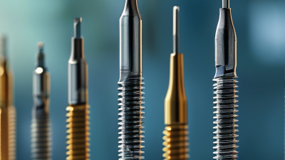
The basic structure includes a threaded shaft, screw head, tip, and drive recess. The shaft enters the bone and provides purchase through its threads. The head contains special threads or shaping that engage the plate hole. The tip may be sharp, tapered, or self-drilling, depending on the design. The drive recess accepts a surgical driver during insertion. Small differences matter.
In clinical practice, surgeons select locking screws according to bone quality, fracture pattern, plate position, and imaging findings. A locking construct can support fragile bone and reduce plate movement. However, it does not automatically create compression across a fracture. That limitation is sometimes overlooked. Conventional screws may still be useful when direct bone-to-plate compression is needed.
Accurate insertion requires correct alignment, controlled torque, and careful attention to the screw length. Excessive force may damage the recess or weaken the connection. A screw that is too long can irritate nearby soft tissue. These details seem basic, yet they influence fixation quality. The best choice depends on the patient, not the screw alone.
How Locking Screws Work Within a Bone Plate System
What Are Orthopedic Locking Screws?
How Locking Screws Work Within a Bone Plate System
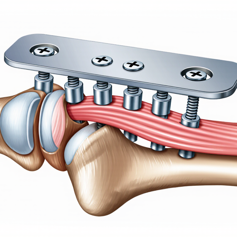
An orthopedic locking screw does more than hold a plate against bone. Its threaded head engages matching threads in the plate hole. This connection creates a fixed-angle relationship between the screw and plate. The plate and screws then act as one stable framework.
The screw head locks into place.
Unlike a conventional screw, a locking screw does not need to press the plate tightly against the bone. This feature can protect the blood supply beneath the plate, especially when surgeons use a bridge-plating technique. The construct supports fractured bone while preserving a controlled space for healing. It can also provide useful stability in softer bone or complex fractures.
Placement still requires precision. The surgeon must select an appropriate screw length, aim along the planned trajectory, and avoid entering a joint. Excessive tightening may damage the screw, plate, or bone. Insufficient engagement can reduce construct stability. The plate must also fit the bone reasonably well, although locking screws cannot correct every contour problem.
The concept sounds simple.
In practice, fracture pattern, bone quality, soft-tissue condition, and patient activity all influence the surgical plan. A locking plate is not automatically stronger in every situation. It may provide limited compression unless paired with suitable techniques or conventional screws. Postoperative imaging and clinical follow-up remain important because fixation can change as bone heals. Even experienced teams must reassess unexpected alignment, pain, or delayed healing rather than rely on the locking mechanism alone.
Common Types and Materials of Locking Screws
What Are Orthopedic Locking Screws?
Common Types and Materials of Locking Screws
Orthopedic locking screws secure into threaded plate holes, creating a stable fixed-angle construct. Unlike standard screws, they do not rely only on the plate pressing against bone. This feature can help maintain alignment near joints or in softer bone.
Common types include fixed-angle, variable-angle, and headless locking screws. Fixed-angle screws follow a predetermined direction. Variable-angle designs allow limited directional adjustment, which can help avoid a fracture line or sensitive anatomy. Headless screws sit beneath the bone surface, making them useful where a raised screw head could irritate nearby tissue. The distinction is not always clean. Screw design must match the fracture pattern, bone quality, and surgical plan.
Stainless steel remains valued for strength, familiarity, and consistent imaging characteristics. Titanium is lighter and generally more compatible with magnetic resonance imaging, although image distortion can still occur. Some systems use titanium alloys for improved strength-to-weight performance. Material choice is not a simple preference. Corrosion behavior, elasticity, fatigue resistance, and future imaging all deserve attention. Small details matter.
Tips: Check the screw diameter, length, thread design, and locking angle before insertion. Confirm the trajectory with fluoroscopy when appropriate. Avoid forcing a screw into a poorly aligned hole. That mistake can damage the thread or reduce fixation quality. Surgeons should follow validated technique guides and assess each patient individually. Tissue handling matters too. A technically strong construct can still fail when biological healing is overlooked.
What Are Orthopedic Locking Screws? Common Types and Materials
Locking screws secure directly into threaded plate holes to create a fixed-angle construct. Common designs include fixed-angle locking screws, variable-angle locking screws, and hybrid locking screws. The chart compares representative elastic modulus values of materials used in orthopedic fixation.
Representative elastic modulus values: 316L stainless steel ≈193 GPa, Ti-6Al-4V titanium alloy ≈114 GPa, cobalt-chromium alloy ≈230 GPa, and PEEK ≈3.6 GPa. Actual values may vary with composition, processing, and test conditions. Most orthopedic locking screws are metallic, especially stainless steel or titanium alloy.
Benefits, Limitations, and Important Surgical Considerations
Orthopedic locking screws secure their threaded heads inside a compatible plate, creating a fixed-angle construct. This design can preserve alignment when bone quality is poor. The International Osteoporosis Foundation reports that one in three women and one in five men over 50 will experience an osteoporotic fracture. That population may benefit from improved fixation stability, especially around the distal radius, proximal humerus, and ankle.
The benefit is not unlimited. A rigid construct can reduce micromotion, yet excessive stiffness may restrict callus formation. Poor plate positioning, inaccurate screw length, or over-tightening can cause joint penetration, soft-tissue irritation, or delayed healing. The implant does not correct weak biology. It only manages mechanics.
Surgical planning matters.
Surgeons should confirm fracture geometry with multiple imaging views and select screw trajectories carefully. Intraoperative fluoroscopy can detect penetration, but it is not infallible. AO Surgery Reference emphasizes correct plate placement, adequate fixation, and protection of surrounding soft tissues. Clinical judgment remains essential. A 2023 review in Injury also noted that locking constructs do not consistently outperform conventional fixation in every fracture pattern. That finding deserves attention. More screws may increase stability, but they can also create an overly rigid bridge. Postoperative radiographs, wound checks, and functional assessment help identify loss of reduction, infection, or delayed union before problems become harder to manage.